
![]()
Watson's and Crick's model for the structure of DNA "has two helical chains each coiled round the same axis." They stated that " each chain consists of phosphate diester groups joining beta-D-deoxyribofuranose residues with 3',5' linkages". Furthermore "both chains follow right-handed helices, but owing to the dyad [Ed: group of two chains] the sequences of the atoms in the two chains run in opposite directions". They go on to state that "the bases are on the inside of the helix and the phosphates on the outside" and that "the structure repeats after 10 residues on each chain".
Proof
To prove the model for the structure of DNA, four questions had to be answered:
Answer to Question 1 What is the shape of the molecule? Is it a straight chain? Are there two or three strands in the helix?
In May of 1952 Franklin obtained the first good X-ray diffraction pattern of the wet B form of DNA. The features of Franklin's photo suggested that DNA had a helical structure based upon the previous work of Stokes and Wilkins and Cochran, Crick, and Vand.
Wilkins is reported to have shown the unpublished X-ray photograph of DNA to Watson and Crick, at which point Watson is reported to have said "The instant I saw the picture my mouth fell open and my pulse began to race.... the black cross of reflections which dominated the picture could arise only from a helical structure... mere inspection of the X-ray picture gave several of the vital helical parameters." Wilkins, Stokes, and Wilson interpreted the results of their X-ray photograph as consistent with a helix of 2 nm diameter. X-ray diffraction studies determined two levels of periodicities along the long axis of the DNA fibers. The primary one had a period of 0.34 nm and the second one had a 3.4 nm period. The X-shaped cross pattern and its symmetry suggested to Franklin and also to Watson and Crick a helix arrangement for the B form of DNA. The Watson and Crick model for DNA called for a double helix. Franklin concluded that the missing fourth layer line agreed with the double helix "if one molecule is displaced from the other by about three-eighths of the fibre-axis period". Thus, the answer to Question 1 came from Franklin's X-ray photo.
Answer to Question 2 Are the chains parallel or anti-parallel? That is do the chains run in the same or opposite directions?
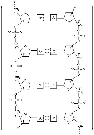
The density data as well as the X-ray data suggest that there are two chains. In the Watson and Crick model, the two chains are held together by a base on one chain linked to a base on the other chain. Adenine and guanine are purines while thymine and cytosine are pyrimidines. A purine on one chain always bonds to a pyrimidine on the other chain. Chargaff's rules suggest that adenine, A, always bonds to thymine, T, and guanine, G, always bonds to cytosine, C, because the percentage of A is always approximately the percentage of T and the percentage of G is always approximately that of C, even though the percentages of A and T may be very different than the percentages of G and C. The purine can be on either chain but its partner pyrimidine must be on the other chain. This suggests that the chains run in opposite directions. The base is attached to carbon 1' of the sugar with phosphates attached to carbons 3' and 5'. In the figure below, the links of the phosphates to the deoxyribose sugars on the left strand run 3' to 5' bottom to top whereas those on the right strand run 3' to 5' top to bottom.
Watson's and Crick's model would suggest that the double helix is "a pair of templates, each of which is complementary to the other", which, prior to duplication, separate and unwind into two chains. "Each chain then acts as a template for the formation on to itself of a new companion chain, so that eventually we shall have two pairs of chains where we only had one before. Franklin's X-ray data also suggested "that there are only two molecules in the primitive unit cell" and that the two chains were offset, which was consistent with the Watson and Crick model. Thus Question 2 is answered.
Answer to Question 3 Are the phosphates on the outside and the bases on the inside of the helix or vice versa?
In her studies Franklin discovered that there were two forms of DNA, a dry, A form, and a wet, B form. The fact that there was a wet B form and a dry A form suggested to Franklin that the phosphates were on the outside and bases on the inside. The wet form could have water molecules attracted by the phosphate groups and sodium counter-ions if the phosphate groups were on the outside. No such attraction would be expected for a structure where the bases were on the outside. Franklin also stated that the features of her X-ray photo suggested that "a crystallographically important part of the molecule lies on a helix of this diameter. This can only be the phosphate groups or phosphorus atoms."
Speaking of their model for the sodium salt of DNA, Watson and Crick stated "As the phosphates are on the outside, cations have easy access to them." Wilkins, Stokes, and Wilson commented that their X-ray photograph "suggests the presence of nitrogen bases arranged like a pile of pennies in the central regions of the helical system". Thus, Question 3 was answered.
Answer to Question 4 How are the bases involved? How many are there per coil (period) of polymer chain? How are they bonded?
In the Watson and Crick model, DNA is composed of two chains joined by complementary bases that are hydrogen bonded to each other. There are 10 residues, nucleotides, in the 3.4 nm period of the chain. The periodicity of 0.34 nm, deduced from the diffraction data, is due to the nucleotide repeat distance. The 3.4 nm layer lines are attributed by Wilkins and Franklin to the repeat distance or period of the chain. This suggested 10 nucleotides per period of chain. These data were the answer to Question 4.
[Early DNA Research] Back | Forward [Annotated Bibliography]