DNA Optical Transform Kit
The DNA Optical Transform Kit assists instructors in teaching students about DNA and how its structure was solved. The kit includes a booklet and the DNA optical transform slide, which is based on an article in the March, 1999 issue of the Journal of Chemical Education by Amand Lucas, et al.
To order a kit contact the Institute
for Chemical Education.
Unfortunately the Institute for Chemical Education at the University of Wisconsin Madison shut down during COVID and has not reopened.
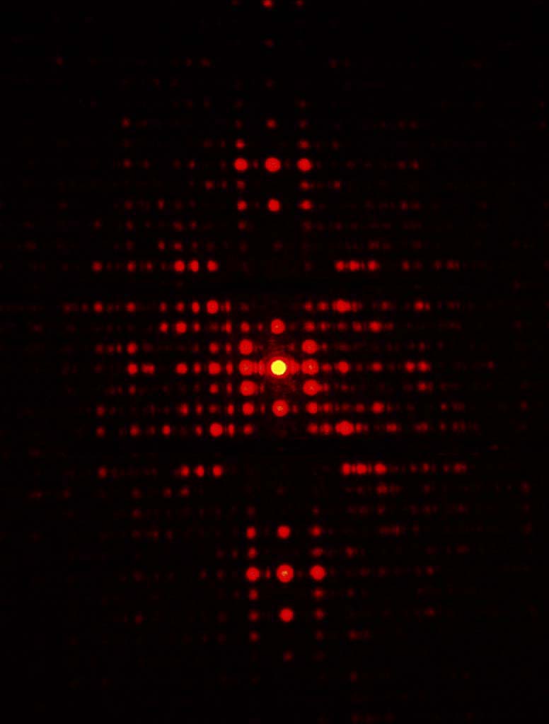
One of the diffraction patterns generated by the DNA optical tranform slide.
The booklet contains:
- a section describing the basic principles of X-ray diffraction that were used to solve the structure of B-DNA;
- a section relating X-ray diffraction to optical transforms;
- a section describing the masks on the DNA optical transform slide and how they relate to the X-ray diffraction structural analysis of DNA;
- an activity that describes how students can use the DNA optical transform slide;
- a copy of the insert, "Liver and Onions: DNA Extraction from Animal and Plant Tissues", from the March, 1999 issue of the Journal of Chemical Education;
- a copy of the article "Revealing the Backbone Structure of B-DNA from Laser Optical Simulations of X-ray Diffraction Diagram" by A. A. Lucas, Ph. Lambin, R. Mairesse, and M. Mathot, which also appears in the March, 1999 issue of the Journal of Chemical Education;
- a section containing a list of publications and web site addresses that describe DNA and the effort to determine its structure; and
- an Appendix containing transparency masters and a suppliers list.
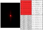
ABCH: These four arrays and their diffraction patterns illustrate how the X-shaped cross in the diffraction image is generated. The sloped lines in arrays B and C result in diffraction patterns of lines that are perpendicular to the arrays: Notice, for example, in B, the lines in the array have positive slope, and the diffraction pattern is a perpendicular line of negative slope. Imagine connecting short positively and negatively sloped lines to form a zig-zag line, which can be smoothed to a sine wave (see array H). The diffraction pattern of array H is an X-shaped cross, which is one of the KEY features of the DNA diffraction pattern.
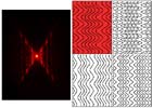
DEHG: These four arrays are all made of sine waves, whose diffraction patterns are all X-shaped crosses. The difference between each of the arrays is the period-to-amplitude ratio of the sine waves. Notice how the X-shaped cross in the diffraction pattern changes as the period-to-amplitude ratio of the sine waves changes in the arrays. By measuring the angles between the arms of the cross in the diffraction pattern, one can calculate the period-to-amplitude ratio in the sine wave, or in the case of DNA, the helix.
HFIL: One of the most distinguishing, and important features of Franklin's DNA diffraction pattern is the missing fourth layer line. If one counts up (or down) from the center of the X-shaped cross, the fourth layer line is absent, or missing in the diffraction pattern. What causes this absence in the diffraction pattern? As is clear in the patterns DEH and G, a single sine wave (or helix) produces an X-shaped diffraction pattern in which there are no missing diffraction lines. This indicates that there must be something more than a single helix in the DNA polymer in order for there to be missing diffraction features. In patterns F, I and L, two sine waves have been overlapped, or offset from one another at different spacings. In F, the offset of the two sine waves is 1/4 of a period, and the second, sixth, and tenth layer lines are missing. In I, the offset is ½ of a period, and every other layer line of diffraction is absent. In L, the offset is 3/8 of a period, and the fourth layer line of diffraction is missing. This is a key piece of information for the understanding of the DNA polymer as a double helix with an offset of the two helices by 3/8 of a period.
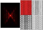
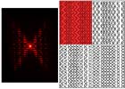
HLKJ: This group of arrays helps illustrate several of the more subtle features of the DNA diffraction pattern. In DNA the phosphorus atoms are part of the sugar-phosphate backbone of the DNA polymer, and, as the heaviest atoms, are the strongest scatterers of X-rays in the experiment. In the diffraction pattern of array K, a pattern of four diamonds is visible. The fact that the north and south diamonds are empty of diffraction intensity, and the east and west diamonds contain diffraction intensity, indicated that the sugar-phosphate backbone was on the outside of the DNA double helix, and the base pairs lie on the inside of the DNA molecule. Lastly, in array J, along with the sine waves of dots, as in array K, ten short horizontal lines have been added to the inside of the double sine waves. These ten horizontal lines represent the ten base pairs per period THAT lie on the inside of the DNA double helix. In the diffraction pattern of J, there are "blobs" (or groups of spots), of diffraction intensity at the top and bottom of the diffraction pattern. These "blobs" can also been seen in Franklin's original DNA diffraction image, and indicate the spacing of the base pairs within the DNA double helix. There are exactly ten base pairs per period of the DNA double helix. This can be confirmed by noting that the "blobs" of diffraction occur at the 10th layer line of the X-shaped cross.
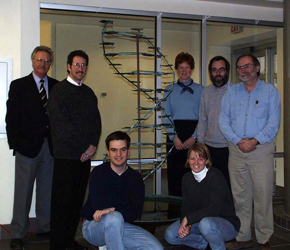
Authors of DNA Optical Transform Kit: standing l-r: Amand Lucas, Art Ellis, Karen Nordell, George Lisensky, Mike Condren; Russ Tobe, and Anne-Marie Jackelen are kneeling.
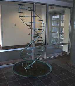
"DNA fountain" in the lobby of Genetics - Biotechnology Building University of Wisconsin - Madison. |
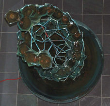
View down the long axis of the "DNA fountain". |

-
This view shows the two hydrogen bonds between thymine and adenine, and the
three hydrogen bonds between cytosine and guanine.